🧠✨ Calling all neuro-enthusiasts!
We’re excited to launch the NSI-ECRN Monthly Microscopy Spotlight — a new initiative to showcase the stunning imaging work of early career neuroscience researchers across Ireland! 🧪🔬
Each month, we’ll feature one standout microscopy image on our socials, celebrating the beauty of brain research and the brilliant scientists behind the lens.
📸 Got an image you’d love to share?
Submit HERE
Let’s highlight the science and the art of neuroscience together.
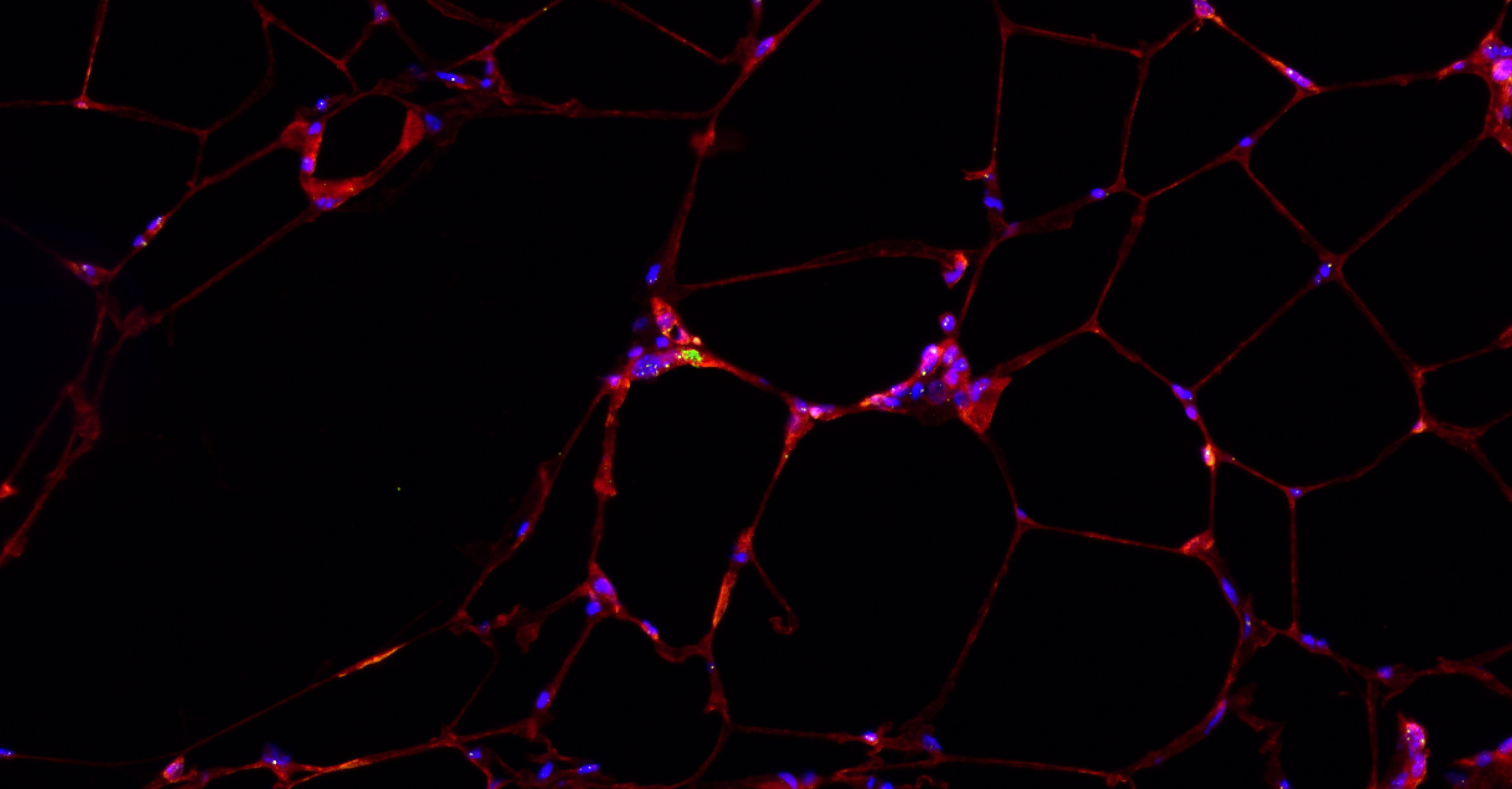
“Fluorescent staining for the macrophage marker Iba1 (red), the inflammatory marker IL-1β (yellow), the cellular stress marker GDF15 (green) and the nuclear marker Dapi (blue) in the adipose tissue of an adult mouse after 12 weeks of high-fat diet feeding and 7 days post-traumatic brain injury, combining IF and FISH (RNAscope).”
-Gideon van der Linden, PhD student in University College Dublin
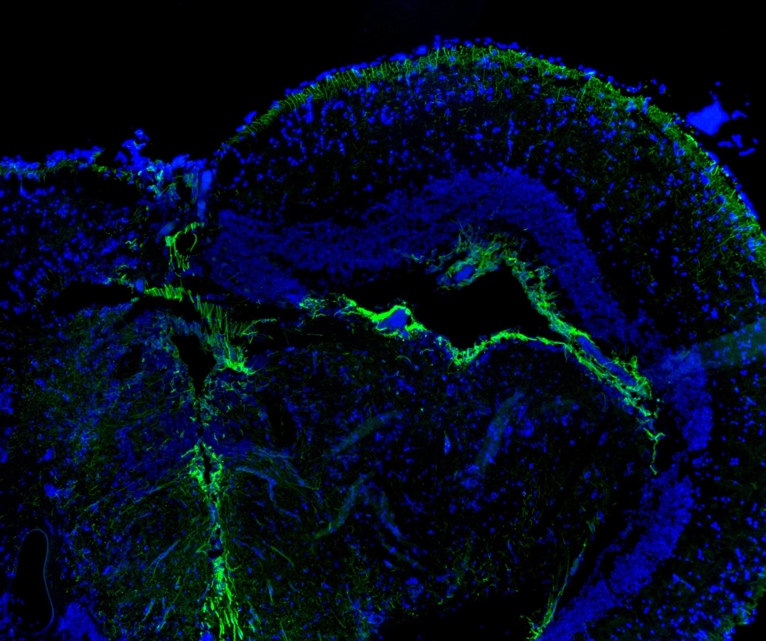
“Male 1 year old Zebrafish coronal midbrain section, probed for GFAP and costained by DAPI”
-Fergus Woods, PhD student in University College Dublin
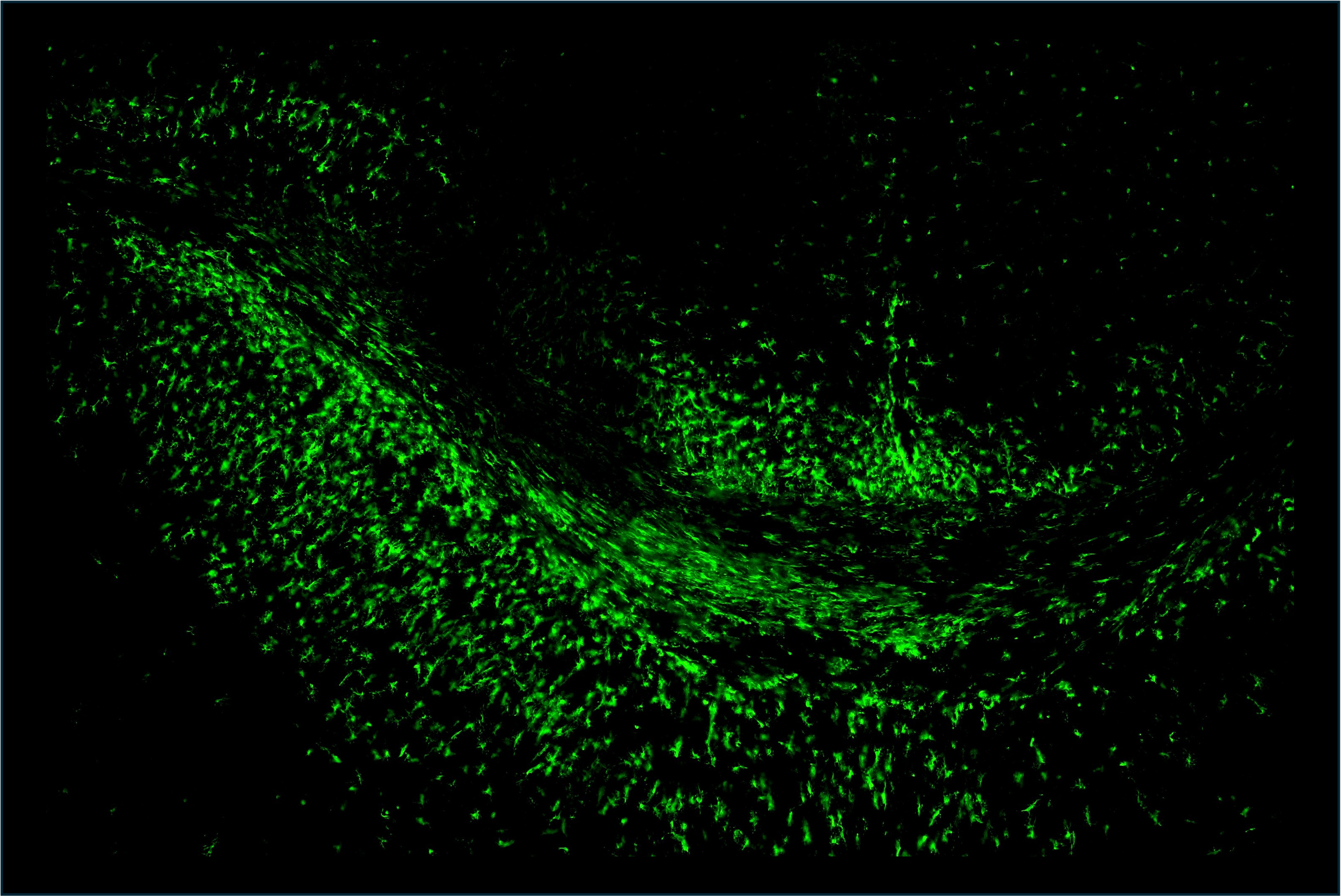
“Microglia hard at work💪🏼✨! An army of microglia infiltrating the corpus callosum region of a Sprague-Dawley rat brain provides a glimpse into the intricate workings of the immune cells of our central nervous system and showcases their essential role in keeping our brains healthy and harmonious.”
-Meike Mulder, Masters student at University College Cork